Healthcare Agency Funding Propels Imaging and AI Research

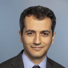
Farzad Fereidouni, Ph.D., recently joined Emory University as an associate professor in the Department of Pathology and Laboratory Medicine. He has extensive expertise in developing imaging instrumentation and computational methods for tissue and cellular microscopy applications. His current focus is on creating novel imaging approaches with direct applications in pathology and patient care, aligning seamlessly with the demands of an effective research program and integration within Emory’s Department of Pathology.
We sat down for an interview regarding how his research meshes with the introduction of Artificial Intelligence technology.
Q: Can you tell us about your recent grant from the National Cancer Institute and its importance to your research?
I received an NCI R01 grant that led me to develop a large-format tissue scanner. This grant, similar to those from ARPA-H, was funded two years ago for about three million dollars. With this funding, we are creating a scanner that can image tissue samples with sizes up to 100x100 mm2 within five minutes after they are received from surgery. This rapid imaging can inform surgeons about positive margins during the procedure. While this project does not use AI components, its development will help integrate AI. The images produced will be digital, making it straightforward to add AI to this creative workflow.
Q: How do you envision the technologies you are developing will use AI to enhance patient accessibility?
I created the terminology of "histology in a box" to refer to the large-format tissue scanner we are developing. Instead of going through large labs to process tissue, our box will receive the tissue and create a digital image that can be analyzed by AI. This approach puts an entire histology lab into a single unit, reducing costs and processing times, and making it more accessible to everyone. On top of that, by enabling AI diagnostics, this system offers real-time, on-site analysis. While the patient is still in bed, all processing and diagnoses can be completed, and the surgeon is notified immediately, improving patient efficiency and accessibility.
Q: What steps need to be taken before your technologies can be integrated with AI solutions?
Before using AI, we need to make sure that we validate our current technology to ensure its accuracy and reliability. About two years ago, we published a study in the Archives of Pathology where we tested our technology with 100 tissue samples from 10 different organs. We compared the results by having five pathologists diagnose the tissues from the images we provided. We then processed these tissues using standard histology processes, creating slides for evaluation.
The study demonstrated a 97% concordance between our technology, which we call slides pre-technology, and standard histology practices. This validation was important as it confirmed the accuracy of our technology and provided a solid foundation for applying it in new research and ARPA-H projects. By proving that our technology aligns with standard histology practices, we have a pinpoint of a reliable basis for further development and integration of AI.